
Page 27 - Delaware Medical Journal - October 2017
P. 27
CASE REPORT
There was hyper pigmentation of the skin overlying the mass without skin thickening. The mass was not tender and was not associated with any nipple discharge, nipple retraction, or axillary lymphadenopathy.
Mammogram showed a 5.5 cm circumscribed round mass in the
upper central right breast as well as
a 2.4 cm oval mass in the lower inner right breast. (Figure 1) Ultrasound showed a 4.1 x 3.9 x 5.9 cm complex cystic and solid mass at the 12 o’clock position 6 cm from the nipple. (Figure 2) There is vascularity to the solid component. There was a 0.7 x 1.8 cm complex cystic and solid component mass communicating with the larger mass, corresponding to mammographic �� � � � � � � �
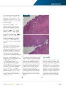
Ultrasound-guided aspiration of the right breast cyst revealed hemorrhagic ����������������������������������� ���������������������������������������� an abscess. Cytology was negative for malignant cells. The patient underwent excision of the cyst two months later after the cyst re-accumulated. Pathology showed invasive ductal carcinoma that was present variably along the entire length of the cavity wall, histology grade 3. (Figure 3) The tumor was
7 cm in maximum diameter. The
cells were estrogen and progesterone receptor negative as well as HER-2 negative. There was no associated Ductal �������������������������������������� The superior surface of the biopsy specimen was discontinuous and tumor involvement in those areas could not
be determined, though within the specimen there was a 1.0 mm inferior and superior margin.
FIGURE 3
The patient then underwent mastectomy with an axillary sentinel node procedure after technetium mapping of her axilla and chest wall. Pathology showed no residual carcinoma in the breast and
the sentinel node was negative. She was staged as a IIB (T3 N0 M0). She met with medical and radiation oncology
to discuss adjuvant chemotherapy, radiation and follow up imaging. After counseling and discussion her therapy options, she decided to forgo radiation and chemotherapy in favor of frequent follow up.
DISCUSSION
Intracystic carcinoma is predominantly reported as a papillary neoplasm. It has been suggested that intracystic papillary carcinoma may be a form of invasive ductal carcinoma or ductal carcinoma in situ.1 Lesions that are localized to cyst ��������������������������������������� intracystic carcinomas, with no reference to extramural or metastatic potential.2 This leads to further speculation on
the pathogenesis of these tumors and ultimately the question of appropriate
Del Med J | October 2017 | Vol. 89 |
No. 10
315
